Within the realm of the clinical Histopathology laboratory (hospital or reference lab), the Hematoxylin & Eosin stain has become a procedural doctrine; validated and written in stone, and rarely, if ever, changed or modified. Typically, this only happens when a decision is made to change from one company’s product to another’s, which is equally rare in H&E staining. Pathologists have personal preferences as to the depth of nuclear stain (hematoxylin), the brilliancy of the counterstain (eosin), and other visual details that they are most comfortable with in making diagnoses. When they achieve the staining results they like, it is rarely changed unless there is a change in pathologist staff.
In the research environment there is more of a project-specific approach where the stain product and protocol are selected and optimized based on variabilities such as:
- tissue type, e.g., brain, bone, cytologic specimens, etc.
- various fixatives used, e.g., bouin’s, zinc, formalin, etc.
- special staining temperature restrictions
Unlike ‘routine’ histology where most things are standardized, the variabilities in research are endless and can change from one project to the next.
When changes from the ‘routine’ have to be made, rather than taking the laborious task of endless multiple staining trials, testing irrelevant steps that make no difference in the actual quality/color/intensity of stain, one should always remember to reduce the number of staining step variables to as few as possible. In H&E staining it can be broken down into 4 major parts: 1) Deparaffinization, 2) Primary & Secondary Dye Staining, 3) Dehydration & Clearing, and 4) Post-Staining Rinses.
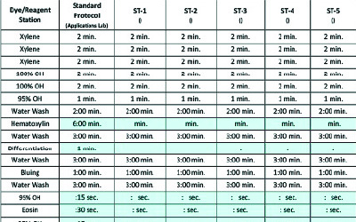
In the Staining Trial Parallels Form (STPF) listed in this article and using the example of formalin-fixed paraffin embedded (FFPE) tissue slides; Deparaffinization, (Part 1) and hydrating slides down to water is a uniform procedure. It does not need to be changed or modified from stain trial to stain trial. Neither does Part 3, the Dehydration & Clearing. The only variables that one needs to deal with are Part 2, Dye Staining, and Part 4, Eosin/Alcohol Post-Staining Rinses.
The STPF shows a number of different staining trials denoted as ST-1, ST-2, etc. Within the form you see set times that do not need to be modified during the various trials. They are illustrated in the clear or white cells. The blue cells demonstrate the only variables that need to be adjusted with each trial until you have optimized your nuclear and cytoplasmic (primary and secondary) staining results. Start with your standard protocol listed by the second column from the left. This would be your baseline from which you will make your subsequent adjustments in staining times. Then systematically adjust the timing steps in the blue cells only, to meet the preference of the Pathologist.
| Dye/Reagent Station |
Standard Protocol (Applications Lab) |
ST-1 () |
ST-2 () |
ST-3 () |
ST-4 () |
ST-5 () |
|---|---|---|---|---|---|---|
| Xylene | 2 min. | 2 min. | 2 min. | 2 min. | 2 min. | 2 min. |
| Xylene | 2 min. | 2 min. | 2 min. | 2 min. | 2 min. | 2 min. |
| Xylene | 2 min. | 2 min. | 2 min. | 2 min. | 2 min. | 2 min. |
| 100% OH | 2 min. | 2 min. | 2 min. | 2 min. | 2 min. | 2 min. |
| 100% OH | 2 min. | 2 min. | 2 min. | 2 min. | 2 min. | 2 min. |
| 95% OH | 1 min. | 1 min. | 1 min. | 1 min. | 1 min. | 1 min. |
| Water Wash | 2:00 min. | 2:00 min. | 2:00 min. | 2:00 min. | 2:00 min. | 2:00 min. |
| Hematoxylin | 6:00 min. | min. | min. | min. | min. | min. |
| Water Wash | 3:00 min. | 3:00 min. | 3:00 min. | 3:00 min. | 3:00 min. | 3:00 min. |
| Differentiation | 1 min. | : | : | : | ||
| Water Wash | 3:00 min. | 3:00 min. | 3:00 min. | 3:00 min. | 3:00 min. | 3:00 min. |
| Bluing | 1:00 min. | 1:00 min. | 1:00 min. | 1:00 min. | 1:00 min. | 1:00 min. |
| Water Wash | 3:00 min. | 3:00 min. | 3:00 min. | 3:00 min. | 3:00 min. | 3:00 min. |
| 95% OH | :15 sec. | : sec. | : sec. | : sec. | : sec. | : sec. |
| Eosin | :30 sec. | : sec. | : sec. | : sec. | : sec. | : sec. |
| 95% OH | :15 sec. | : sec. | : sec. | : sec. | : sec. | : sec. |
| 100% OH | :30 sec. | :30 sec. | :30 sec. | :30 sec. | :30 sec. | :30 sec. |
| 100% OH | 1:00 min. | 1:00 min. | 1:00 min. | 1:00 min. | 1:00 min. | 1:00 min. |
| 100% OH | 2:00 min | 2:00 min | 2:00 min | 2:00 min | 2:00 min | 2:00 min |
| Xylene | 2:00 min. | 2:00 min. | 2:00 min. | 2:00 min. | 2:00 min. | 2:00 min. |
| Xylene | 2:00 min. | 2:00 min. | 2:00 min. | 2:00 min. | 2:00 min. | 2:00 min. |
One must understand and appreciate that water, even on 5% (resulting in 95%), will slowly bleed the eosin dye back out of the tissue. Prolonged time in this post dye step can defeat the purpose of whatever time you set for Eosin, because it will either come back out in the rinse, or worse, slowly bleed out after the slides have been coverslipped.
In using the STPF model, one can simply and systematically complete this process in 3-5 staining runs. It is a perfect model for the research environment where variability and change is in many cases the norm. It is equally, although not as prevalent, useful to the clinical lab environment, because there are always new improvements on staining dye contents as well as protocol. Years ago, laboratories only had the option of using a progressive or regressive method of performing the H&E stain. However, we now have options of using hematoxylins with little to know oxidation, requiring less changes and filtering of precipitates. We have options to differentiation solutions where instead of just using a harsh acid-alcohol reagent, we can use a milder acetic acid reagent. Eosins have a stronger dye content and additives (as previously mentioned) that give a more vibrant coloration. Thus, we now have hybrid staining methods that can give us an even greater intensity in our H&E protocol. The science and the chemistries have expanded. The method of optimizing your stain has been presented. What’s left is getting your Pathologist to consider an opportunity to make his/her job easier. Use the STPF model and experiment with different variables in your own time. Then one day surprise your Pathologist with a few stain trials to review and consider. It is our job as Histology professionals to continue to adapt and develop improved methods for doctors, not to change, but to consider. Good luck!!!
References
- Brown, S., ‘Slide Match Protocol’, Lab Management Consultants, Technical Series, 2010.
- Brown, S., “A Systems Approach to H&E Staining”, Workshop presented to the American Society for Clinical Pathologists (ASCP), 2019