Herbert Skip Brown, M. Div. HT(ASCP)
Lab Storage Systems, Inc.
The rotary microtome is the central core of clinical histopathology instrumentation; and even though it has been modernized from manual to semi and fully automated units, basic manual hygiene and maintenance is imperative to maintain the ability to optimally section tissue specimen. Failure to perform simple basic functions on a regular basis will eventually result in microtomy artifacts such as inconsistent ribboning, thick and thin sections, and a host of other challenges including total operational failure of the microtome unit.
As microtome design has transitioned over time, so has the maintenance needs of the unit. Once it was the responsibility of the user to do certain preventative maintenance tasks such as oiling the gears, lubricating the rotary wheel, etc.; as with the authors first unit used in the 1970s, the American Optical (AO 820) (Left).
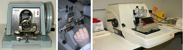
With this unit the housing lid would lift up and the user had access to all the gears and tension wires. Eventually this transitioned to microtomes that had a specific single port hole for oil, as in the Leitz 1520 microtome in the 1980s (Center). The inner mechanism was not accessible to the user. Eventually modernization and technology took the task of oiling as part of maintenance away from the user and was done only through trained service technicians. An example of this was the Leica RM 2235 (Right). All microtomes were optimally designed with precision manufacture and were exceptional for their time and need.
One common aspect of all three examples was the need for the user to thoroughly clean the unit after each day or event of use. While the inner mechanism was no longer accessible, the knife or blade holder still required personal attention consisting of complete removal of all paraffin shavings and debris. Likewise, the entire workstation including countertop and floor required daily cleaning and hygiene. With respect to the blade holder, paraffin will cake up on the surface and in the crevices of the holder.
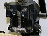
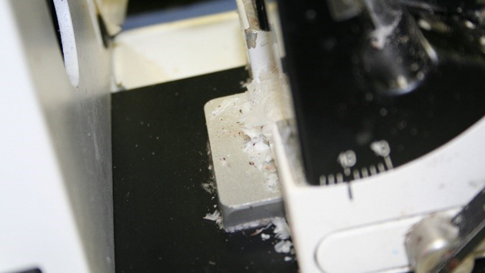
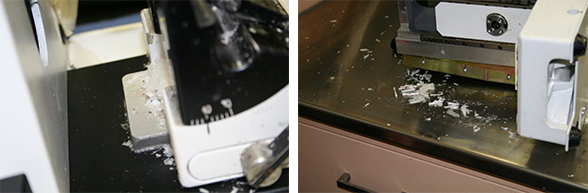
Also, the chuck which holds the block can become caked with fragments, debris, and even chunks from previous tissue blocks sectioned, clogging the chuck and impeding it from tightly securing the block resulting in thick and thin sections. The locking mechanism on the holder and chuck can also become clogged and keep the block from positioning correctly and holding firm. Both must be thoroughly cleaned after use as well as the surface of the microtome and the waste tray. Removal of the blade holder from the microtome is necessary for a complete and thorough cleaning. The image above demonstrates lack of proper cleaning where paraffin debris is trapped behind the blade holder (Left image). This is why proper cleaning requires removal of the holder.
With respect to the workstation, as previously mentioned, all immediate countertop areas and floor space around the microtome should be cleaned with each user or at least daily. Bacteria and germs from tissue debris and waste can build up and create a personal safety issue and general basic hygiene concerns. The right image shows excessive paraffin shavings and debris around and under the microtome on the countertop. These are prime areas that are often left not cleaned where build up is not easily noticeable.
Daily cleaning must be a commitment of all users. This can easily be achieved with a brush for the visible particles, followed by the use of gauze dampened with xylene and wiping down the microtome unit, blade holder, and workstation. Residual xylene is then removed by dampening gauze with 100% alcohol and going over the same areas. Some technicians have gone so far as to totally immerse their blade holder in xylene. This is strongly not advisable, as many of the knife holder have hard rubber bushings internally to help secure the settings. Xylene can/will affect these bushings and cause them to lose their strength. Paraffin cleaning sprays have been developed by commercial vendors, but many equipment repair companies advise caution when using these. When using this, paraffin can melt on the knife holder and trickle down into the crevices of the holder. After time there is a build-up of paraffin inside the holder because it has not been removed; only melted down and re-solidified internally in the holder. This will eventually build up and impede the holder from being able to secure the blade tightly.
For the immediate floor area, a scraper should be first used, with the debris swept up and discarded. In many high-volume laboratories where microtomy is done on more than one shift, most institutions dont have the luxury or funding to have a personal microtome and workstation for each user; and therefore, one microtome may have multiple users in a 24-hour period. To ensure compliance with these standards, supervisors should write the cleaning and maintenance procedures into their SOP section on microtomy. Daily walk-by inspection of all cutting stations should also be considered so that failure to adhere to these standards can be immediately addressed. Supervisors should also incorporate a regular preventative maintenance cleaning from trained service repair representatives, or a trained bio-med technician at their institution. This should be done every 6-12 months annually.
As previously mentioned, it takes a personal commitment from each user to follow these basic preventative maintenance steps. Only then can we extend the life span of microtome units, and ensure optimal working perfection in tissue sectioning.